You have no items in your shopping cart.
Description
Research Area
Product Categories/Products/Antibodies,Product Categories/Research Area,Cancer,Product Categories/Products/Antibodies/Primary Antibodies,Epigenetics
Images & Validation
−Item 1 of 7
| Tested Applications | IF, IHC-P, WB |
|---|---|
| Dilution Range | Western blot: 1-2ug/ml,Immunofluorescence: 1-2ug/ml,Immunohistochemistry (FFPE): 1-2ug/ml |
| Reactivity | Human, Mouse |
| Application Notes |
Key Properties
−| Antibody Type | Primary Antibody |
|---|---|
| Host | Mouse |
| Clonality | Monoclonal |
| Isotype | Mouse IgG1, kappa |
| Clone No. | BMI1/2823 |
| Immunogen | A recombinant human partial protein (amino acids 142-326) was used as the immunogen for this BMI1 antibody. |
| Purity | Protein G affinity chromatography |
| Conjugation | Unconjugated |
Storage & Handling
−| Storage | Store the BMI1 antibody at 2-8°C (with azide) or aliquot and store at -20°C or colder (without azide). |
|---|---|
| Buffer/Preservatives | 0.2 mg/ml with 0.1 mg/ml rAlbumin (US sourced), 0.05% sodium azide |
| Expiration Date | 12 months from date of receipt. |
| Hazard Information | This BMI1 antibody is available for research use only. |
| Disclaimer | For research use only |
Similar Products
−- Item 1 of 8
BMI1 antibody [orb19786]
ICC, IF, IHC-P, WB
Guinea pig, Human, Mouse, Rat
Rabbit
Polyclonal
Unconjugated
100 μg - Item 1 of 7
- Item 1 of 7
- Item 1 of 7
- Item 1 of 7


Quality Guarantee
Explore bioreagents carefree to elevate your research. All our products are rigorously tested for performance. If a product does not perform as described on its datasheet, our scientific support team will provide expert troubleshooting, a prompt replacement, or a refund. For full details, please see our Terms & Conditions and Buying Guide. Contact us at [email protected].

Western blot testing of mouse NIH3T3 cell lysate with BMI1 antibody. Predicted molecular weight: 37-43 kDa.

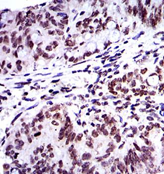
IHC staining of FFPE human colon carcinoma with BMI1 antibody. HIER: boil tissue sections in pH9 10mM Tris with 1mM EDTA for 20 min and allow to cool before testing.

IHC staining of FFPE human breast carcinoma with BMI1 antibody. HIER: boil tissue sections in pH9 10mM Tris with 1mM EDTA for 20 min and allow to cool before testing.

IHC staining of FFPE human prostate carcinoma with BMI1 antibody. HIER: boil tissue sections in pH9 10mM Tris with 1mM EDTA for 20 min and allow to cool before testing.

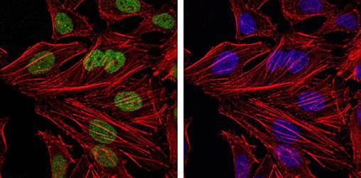
Immunofluorescent staining of PFA-fixed human HeLa cells with BMI1 antibody (green) and Phalloidin (red).

Analysis of HuProt (TM) microarray containing more than 19000 full-length human proteins using BMI1 antibody (clone BMI1/2823). These results demonstrate the foremost specificity of the BMI1/2823 mAb. Z- and S- score: The Z-score represents the strength of a signal that an antibody (in combination with a fluorescently-tagged anti-IgG secondary Ab) produces when binding to a particular protein on the HuProt (TM) array. Z-scores are described in units of standard deviations (SD's) above the mean value of all signals generated on that array. If the targets on the HuProt (TM) are arranged in descending order of the Z-score, the S-score is the difference (also in units of SD's) between the Z-scores. The S-score therefore represents the relative target specificity of an Ab to its intended target.

SDS-PAGE analysis of purified, BSA-free BMI1 antibody as confirmation of integrity and purity.
Quick Database Links
UniProt
UniProt Details
− No UniProt data available
Documents Download
Datasheet
Product Information
Request a Document
Protocol Information
WB
Western Blot (IB, immunoblot)
IHC-P
Immunohistochemistry Paraffin
IF
Immunofluorescence
BMI1 Antibody (orb639924)

- 0.0
Based on 0 reviews
Participating in our Biorbyt product reviews program enables you to support fellow scientists by sharing your firsthand experience with our products.
Login to Submit a ReviewAvailable Sizes
Select a size below
Free Secondary Antibody (20 ul)0/0
Please add an antibody product to your cart first.