You have no items in your shopping cart.
Featured
Description
Research Area
,Cancer,Cell Cycle,Neuroscience,Signal Transduction
Images & Validation
−Item 1 of 4
| Tested Applications | FC, IF, IHC-P, WB |
|---|---|
| Reactivity | Human |
| Application Notes |
Key Properties
−| Antibody Type | Primary Antibody |
|---|---|
| Clonality | Polyclonal |
| Isotype | Rabbit Ig |
| Immunogen | This FGFR4 antibody is generated from rabbits immunized with a KLH conjugated synthetic peptide between 24-55 amino acids from the N-terminal region of human FGFR4. |
| Target | FGFR4 |
| Molecular Weight | 88 kDa |
| Purification | This antibody is prepared by Saturated Ammonium Sulfate (SAS) precipitation followed by dialysis |
| Conjugation | Unconjugated |
Storage & Handling
−| Storage | Maintain refrigerated at 2-8°C for up to 2 weeks. For long term storage store at -20°C in small aliquots to prevent freeze-thaw cycles. |
|---|---|
| Form/Appearance | Liquid |
| Buffer/Preservatives | Supplied in PBS with 0.09% (W/V) sodium azide. |
| Concentration | batch dependent |
| Expiration Date | 12 months from date of receipt. |
| Disclaimer | For research use only |
Alternative Names
−Fibroblast growth factor receptor 4, FGFR-4, CD334, FGFR4, JTK2, TKF
Similar Products
−- Item 1 of 1
Human Fibroblast Growth Factor Receptor 4 (FGFR4) ELISA Kit [orb777025]
Human
0.16-10 ng/mL
0.066 ng/mL
48 T, 96 T - Item 1 of 1
Rat Fibroblast Growth Factor Receptor 4 (FGFR4) ELISA Kit [orb1817334]
Rat
0.32-20 ng/mL
0.11 ng/mL
48 T, 96 T - Item 1 of 5
- Item 1 of 5
- Item 1 of 3
Anti-FGFR4 / CD334 Reference Antibody (U3-1784) [orb1806211]
ELISA, FA, FACS, Kinetics
Human
Monoclonal
Unconjugated
100 μg, 1 mg, 5 mg, 50 μg


Quality Guarantee
Explore bioreagents carefree to elevate your research. All our products are rigorously tested for performance. If a product does not perform as described on its datasheet, our scientific support team will provide expert troubleshooting, a prompt replacement, or a refund. For full details, please see our Terms & Conditions and Buying Guide. Contact us at [email protected].
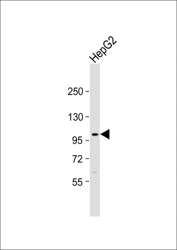
Western Blot at 1:1000 dilution + HepG2 whole cell lysate Lysates/proteins at 20 ug per lane.
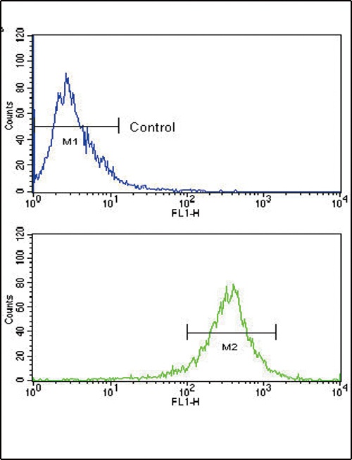
Flow cytometric analysis of WiDr cells using FGFR4 Antibody (bottom histogram) compared to a negative control (top histogram). FITC-conjugated goat-anti-rabbit secondary antibodies were used for the analysis.
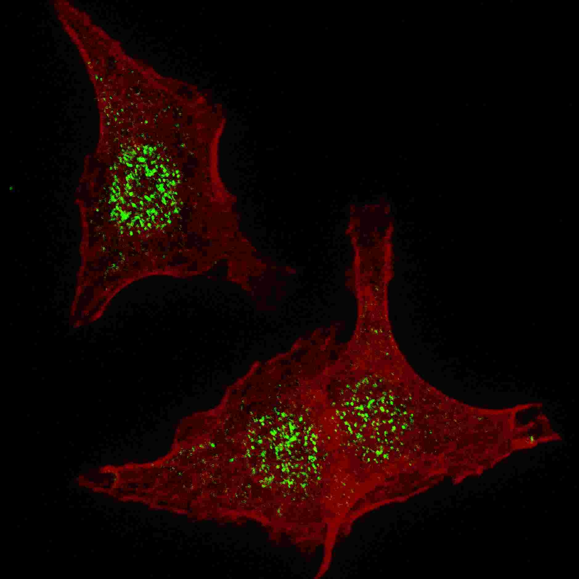
Fluorescent confocal image of HeLa cells stained with FGFR4 antibody. HeLa cells were fixed with 4% PFA (20 min), permeabilized with Triton X-100 (0.2%, 30 min). Cells were then incubated with FGFR4 primary antibody (1:200, 2 h at room temperature). For secondary antibody, Alexa Fluor 488 conjugated donkey anti-rabbit antibody (green) was used (1:1000, 1h). Nuclei were counterstained with Hoechst 33342 (blue) (10 ug/ml, 5 min). Note the highly specific localization of the FGFR4 mainly to the nucleus, supported by Human Protein Atlas
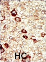
Formalin-fixed and paraffin-embedded human cancer tissue reacted with the primary antibody, which was peroxidase-conjugated to the secondary antibody, followed by AEC staining. BC = breast carcinoma; HC = hepatocarcinoma.
Documents Download
Datasheet
Product Information
Request a Document
Protocol Information
WB
Western Blot (IB, immunoblot)
IHC-P
Immunohistochemistry Paraffin
FC
Flow Cytometry
IF
Immunofluorescence
FGFR4 Antibody (orb1271018)

- 0.0
Based on 0 reviews
Participating in our Biorbyt product reviews program enables you to support fellow scientists by sharing your firsthand experience with our products.
Login to Submit a ReviewAvailable Sizes
Select a size below
Free Secondary Antibody (20 ul)0/0
Please add an antibody product to your cart first.