You have no items in your shopping cart.
Featured
Description
Research Area
,Signal Transduction
Images & Validation
−Item 1 of 3
| Tested Applications | IF, WB |
|---|---|
| Reactivity | Human |
| Predicted Reactivity | Bovine, Gallus, Hamster, Mouse, Rat |
| Application Notes |
Key Properties
−| Antibody Type | Primary Antibody |
|---|---|
| Clonality | Polyclonal |
| Isotype | Rabbit Ig |
| Immunogen | This HSPD1 antibody is generated from rabbits immunized with a KLH conjugated synthetic peptide between 187-215 amino acids from the Central region of human HSPD1. |
| Target | HSPD1 |
| Molecular Weight | 61 kDa |
| Purification | This antibody is prepared by Saturated Ammonium Sulfate (SAS) precipitation followed by dialysis |
| Conjugation | Unconjugated |
Storage & Handling
−| Storage | Maintain refrigerated at 2-8°C for up to 2 weeks. For long term storage store at -20°C in small aliquots to prevent freeze-thaw cycles. |
|---|---|
| Form/Appearance | Liquid |
| Buffer/Preservatives | Supplied in PBS with 0.09% (W/V) sodium azide. |
| Concentration | batch dependent |
| Expiration Date | 12 months from date of receipt. |
| Disclaimer | For research use only |
Alternative Names
−60 kDa heat shock protein, mitochondrial, 60 kDa chaperonin, Chaperonin 60, CPN60, Heat shock protein 60, HSP-60, Hsp60, HuCHA60, Mitochondrial matrix protein P1, P60 lymphocyte protein, HSPD1, HSP60
Similar Products
−- Item 1 of 9
Hsp60/HSPD1 Antibody (monoclonal, 6G2) [orb570314]
FC, ICC, IF, IHC, WB
Human, Mouse, Rat
Mouse
Monoclonal
Unconjugated
100 μg - Item 1 of 5
HSP60 Rabbit Polyclonal Antibody [orb10846]
ICC, IF, IHC-Fr, IHC-P, WB
Bovine, Canine, Equine, Rabbit
Human, Mouse, Rat
Rabbit
Polyclonal
Unconjugated
50 μl, 100 μl, 200 μl - Item 1 of 6
- Item 1 of 6
HSP60 Antibody [LK1] [orb1252759]
FC, IF, IHC, WB
Bovine, Canine, Drosophila, Frog, Gallus, Hamster, Human, Monkey, Mouse, Porcine, Rabbit, Rat, Sheep
Monoclonal
Unconjugated
100 μg - Item 1 of 4
HSP60/HSPD1 (aa333-344) Antibody [orb334054]
ELISA, IF, IHC, WB
Bovine, Canine
Human, Porcine, Rat
Goat
Polyclonal
Unconjugated
100 μg


![HSP60 Antibody [LK1]](/images/pub/media/catalog/product/NewWebsite/15/orb1252759_1.jpg)
![HSP60 Antibody [LK1]](/images/pub/media/catalog/product/NewWebsite/15/orb1252759_2.jpg)
![HSP60 Antibody [LK1]](/images/pub/media/catalog/product/NewWebsite/15/orb1252759_3.jpg)
![HSP60 Antibody [LK1]](/images/pub/media/catalog/product/NewWebsite/15/orb1252759_4.jpg)
![HSP60 Antibody [LK1]](/images/pub/media/catalog/product/NewWebsite/15/orb1252759_5.jpg)
![HSP60 Antibody [LK1]](/images/pub/media/catalog/product/NewWebsite/15/orb1252759_6.jpg)
Quality Guarantee
Explore bioreagents carefree to elevate your research. All our products are rigorously tested for performance. If a product does not perform as described on its datasheet, our scientific support team will provide expert troubleshooting, a prompt replacement, or a refund. For full details, please see our Terms & Conditions and Buying Guide. Contact us at [email protected].
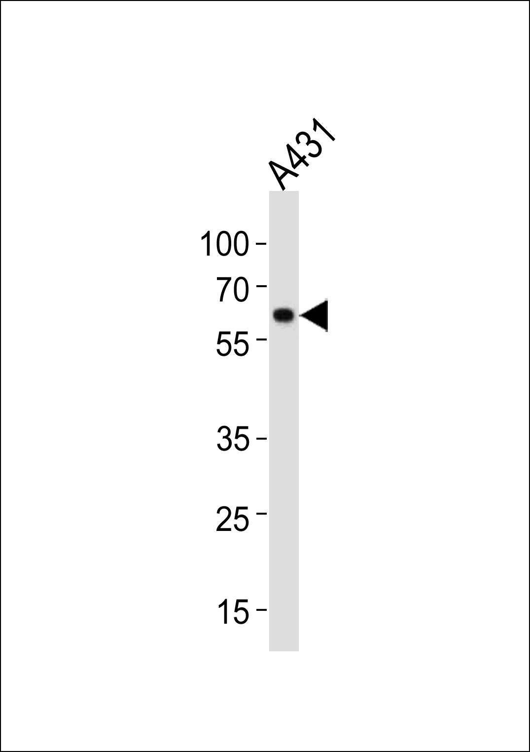
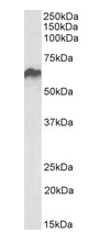
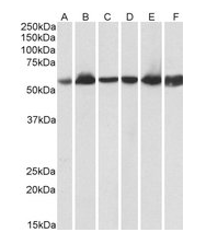
Western blot analysis in A431 cell line lysates (35 ug/lane).
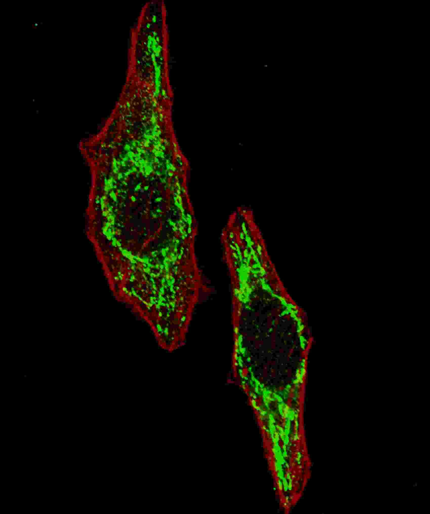
Fluorescent image of U251 cells stained with HSPD1 antibody. U251 cells were fixed with 4% PFA (20 min), permeabilized with Triton X-100 (0.2%, 30 min). Cells were then incubated with HSPD1 primary antibody (1:100, 2 h at room temperature). For secondary antibody, Alexa Fluor 488 conjugated donkey anti-rabbit antibody (green) was used (1:1000, 1h). Cytoplasmic actin was counterstained with Alexa Fluor 555 (red) conjugated Phalloidin (5.25 uM, 25 min). Pictures were taken on a Biorevo microscope (BZ-900, Keyence). Note the highly specific localization of the HSPD1 mainly to the mitochondria, supported by Human Protein Atlas Data (http://www.proteinatlas.org/ENSG00000144381).
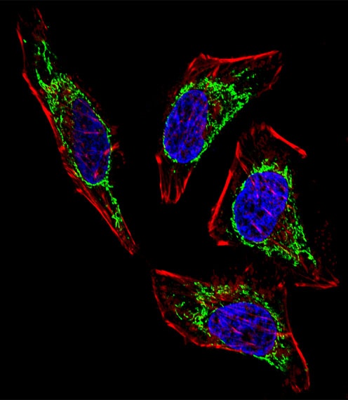
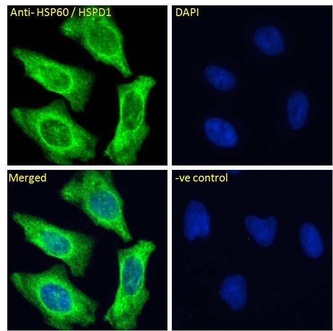
Fluorescent confocal image of Hela cell stained with HSPD1 Antibody. Hela cells were fixed with 4% PFA (20 min), permeabilized with Triton X-100 (0.1%, 10 min), then incubated with HSPD1 primary antibody (1:25). For secondary antibody, Alexa Fluor 488 conjugated donkey anti-rabbit antibody (green) was used (1:400). Cytoplasmic actin was counterstained with Alexa Fluor 555 (red) conjugated Phalloidin (7 units/ml). Nuclei were counterstained with DAPI (blue) (10 ug/ml, 10 min). HSPD1 immunoreactivity is localized to Mitochondria significantly.
Documents Download
Datasheet
Product Information
Request a Document
Protocol Information
WB
Western Blot (IB, immunoblot)
IF
Immunofluorescence
HSPD1 Antibody (orb1264552)

- 0.0
Based on 0 reviews
Participating in our Biorbyt product reviews program enables you to support fellow scientists by sharing your firsthand experience with our products.
Login to Submit a ReviewAvailable Sizes
Select a size below
Free Secondary Antibody (20 ul)0/0
Please add an antibody product to your cart first.