You have no items in your shopping cart.
Featured
Description
Research Area
,Signal Transduction
Images & Validation
−Item 1 of 5
| Tested Applications | FC, IHC-P, WB |
|---|---|
| Reactivity | Human, Mouse, Rat |
| Application Notes |
Key Properties
−| Antibody Type | Primary Antibody |
|---|---|
| Clonality | Polyclonal |
| Isotype | Rabbit Ig |
| Immunogen | This RAB7 antibody is generated from rabbits immunized with a KLH conjugated synthetic peptide between 176-204 amino acids from the C-terminal region of human RAB7. |
| Target | RAB7A |
| Molecular Weight | 23 kDa |
| Purification | This antibody is purified through a protein A column, followed by peptide affinity purification. |
| Conjugation | Unconjugated |
Storage & Handling
−| Storage | Maintain refrigerated at 2-8°C for up to 2 weeks. For long term storage store at -20°C in small aliquots to prevent freeze-thaw cycles. |
|---|---|
| Form/Appearance | Liquid |
| Buffer/Preservatives | Supplied in PBS with 0.09% (W/V) sodium azide. |
| Concentration | batch dependent |
| Expiration Date | 12 months from date of receipt. |
| Disclaimer | For research use only |
Alternative Names
−Ras-related protein Rab-7a, RAB7A, RAB7
Similar Products
−- Item 1 of 21
Anti-RAB7/RAB7A Antibody [orb1676540]
ELISA, FC, IHC, WB
Human, Mouse, Rat
Rabbit
Polyclonal
Unconjugated
10 μg, 100 μg - Item 1 of 7
RAB7 Antibody (C-term) [orb1928362]
FC, IHC-P, WB
Human, Mouse, Rat
Rabbit
Polyclonal
Unconjugated
400 μl - Item 1 of 7
- Item 1 of 1
Human RAB7A, Member RAS Oncogene Family (RAB7A) ELISA Kit [orb779010]
Human
0.32-20 ng/mL
0.112 ng/mL
96 T, 48 T - Item 1 of 3


Quality Guarantee
Explore bioreagents carefree to elevate your research. All our products are rigorously tested for performance. If a product does not perform as described on its datasheet, our scientific support team will provide expert troubleshooting, a prompt replacement, or a refund. For full details, please see our Terms & Conditions and Buying Guide. Contact us at [email protected].
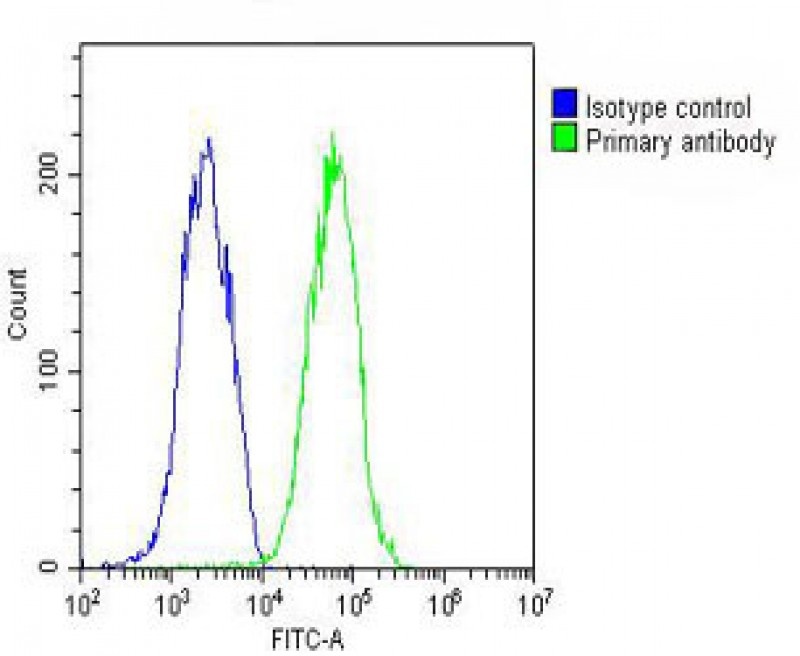
Overlay histogram showing HepG2 cells stained with Antibody (green line). The cells were fixed with 2% paraformaldehyde (10 min) and then permeabilized with 90% methanol for 10 min. The cells were then icubated in 2% bovine serum albumin to block non-specific protein-protein interactions followed by the antibody (1:25 dilution) for 60 min at 37°C. The secondary antibody used was Goat-Anti-Rabbit IgG, Conjugated Highly Cross-Adsorbed at 1/200 dilution for 40 min at 37°C. Isotype control antibody (blue line) was rabbit IgG (1ug/1x10^6 cells) used under the same conditions. Acquisition of > 10000 events was performed.
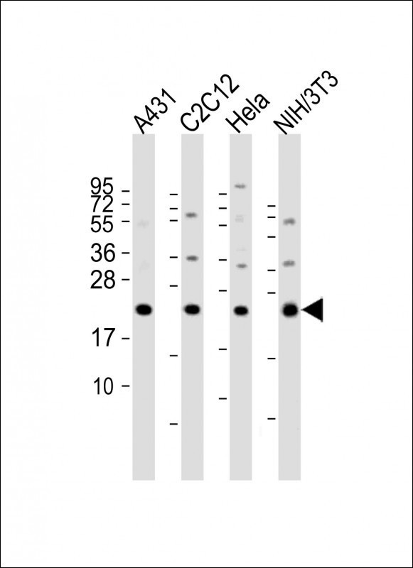
Western Blot at 1:2000 dilution Lane 1: A431 whole cell lysate Lane 2: C2C12 whole cell lysate Lane 3: Hela whole cell lysate Lane 4: NIH/3T3 whole cell lysate Lysates/proteins at 20 ug per lane.
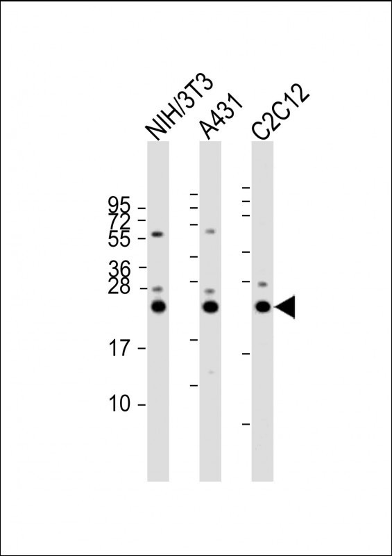
Western Blot at 1:2000 dilution Lane 1: NIH/3T3 whole cell lysate Lane 2: A431 whole cell lysate Lane 3: C2C12 whole cell lysate Lysates/proteins at 20 ug per lane.
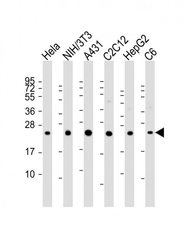
Western Blot at 1:2000 dilution Lane 1: Hela whole cell lysate Lane 2: NIH/3T3 whole cell lysate Lane 3: A431 whole cell lysate Lane 4: C2C12 whole cell lysate Lane 5: HepG2 whole cell lysate Lane 6: C6 whole cell lysate Lysates/proteins at 20 ug per lane.
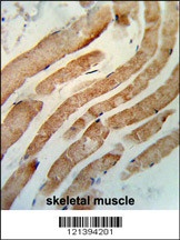
Formalin-fixed and paraffin-embedded human skeletal muscle reacted with RAB7 Antibody, which was peroxidase-conjugated to the secondary antibody, followed by DAB staining.
Documents Download
Datasheet
Product Information
Request a Document
Protocol Information
WB
Western Blot (IB, immunoblot)
IHC-P
Immunohistochemistry Paraffin
FC
Flow Cytometry
RAB7 Antibody (orb1262576)

- 0.0
Based on 0 reviews
Participating in our Biorbyt product reviews program enables you to support fellow scientists by sharing your firsthand experience with our products.
Login to Submit a ReviewAvailable Sizes
Select a size below
Free Secondary Antibody (20 ul)0/0
Please add an antibody product to your cart first.