You have no items in your shopping cart.
Featured
Description
Research Area
,Obesity,Signal Transduction
Images & Validation
−Item 1 of 6
| Tested Applications | FC, IF, IHC-P, WB |
|---|---|
| Reactivity | Human |
| Application Notes |
Key Properties
−| Antibody Type | Primary Antibody |
|---|---|
| Clonality | Polyclonal |
| Isotype | Rabbit Ig |
| Immunogen | This TFAM antibody is generated from rabbits immunized with a KLH conjugated synthetic peptide between 216-246 amino acids from the C-terminal region of human TFAM. |
| Target | TFAM |
| Molecular Weight | 29 kDa |
| Purification | This antibody is purified through a protein A column, followed by peptide affinity purification. |
| Conjugation | Unconjugated |
Storage & Handling
−| Storage | Maintain refrigerated at 2-8°C for up to 2 weeks. For long term storage store at -20°C in small aliquots to prevent freeze-thaw cycles. |
|---|---|
| Form/Appearance | Liquid |
| Buffer/Preservatives | Supplied in PBS with 0.09% (W/V) sodium azide. |
| Concentration | batch dependent |
| Expiration Date | 12 months from date of receipt. |
| Disclaimer | For research use only |
Alternative Names
−Transcription factor A, mitochondrial, mtTFA, Mitochondrial transcription factor 1, MtTF1, Transcription factor 6, TCF-6, Transcription factor 6-like 2, TFAM, TCF6, TCF6L2
Similar Products
−- Item 1 of 10
- Item 1 of 6
- Item 1 of 6
- Item 1 of 1
Human Transcription Factor A, Mitochondrial (TFAM) ELISA Kit [orb777320]
Human
0.32-20 ng/mL
0.118 ng/mL
96 T, 48 T - Item 1 of 1
Mouse Transcription Factor A, Mitochondrial (TFAM) ELISA Kit [orb780878]
Mouse
0.32-20 ng/mL
0.116 ng/mL
96 T, 48 T


Quality Guarantee
Explore bioreagents carefree to elevate your research. All our products are rigorously tested for performance. If a product does not perform as described on its datasheet, our scientific support team will provide expert troubleshooting, a prompt replacement, or a refund. For full details, please see our Terms & Conditions and Buying Guide. Contact us at [email protected].
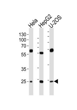
Western blot analysis of lysates from Hela, HepG2, U-2OS cell line (from left to right), using TFAM Antibody at 1:1000 at each lane.
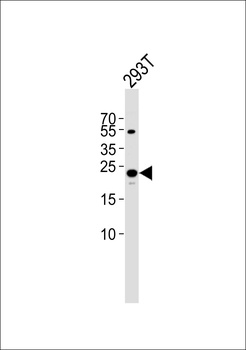
Western blot analysis of lysate from 293T cell line, using TFAM Antibody at 1:1000.
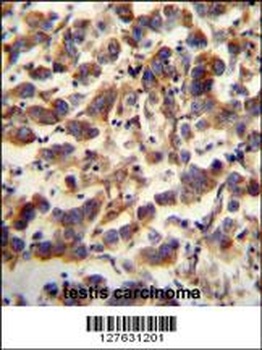
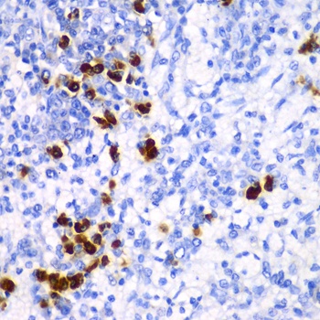
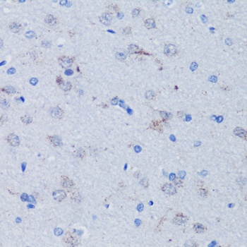
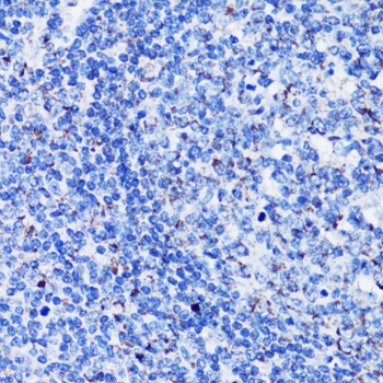
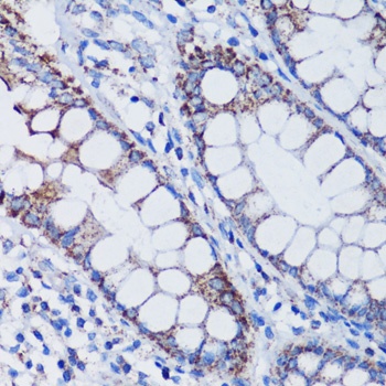
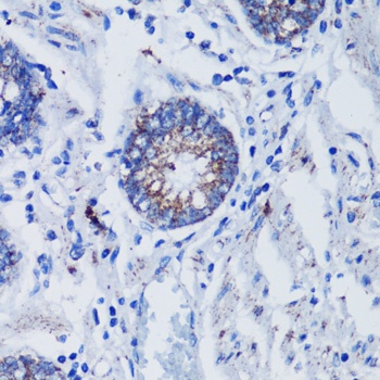
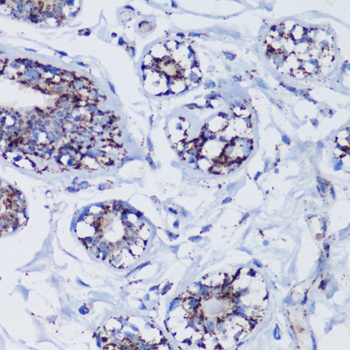
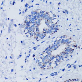
TFAM antibody immunohistochemistry analysis in formalin fixed and paraffin embedded human testis carcinoma followed by peroxidase conjugation of the secondary antibody and DAB staining.
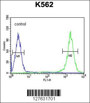
Flow cytometric analysis of K562 cells (right histogram) compared to a negative control cell (left histogram). FITC-conjugated goat-anti-rabbit secondary antibodies were used for the analysis.
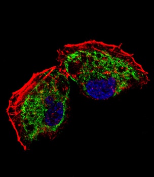
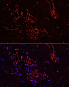
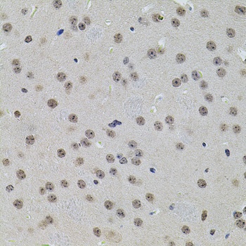
Fluorescent confocal image of NCI-H460 cell stained with TFAM Antibody. NCI-H460 cells were fixed with 4% PFA (20 min), permeabilized with Triton X-100 (0.1%, 10 min), then incubated with TFAM primary antibody (1:25). For secondary antibody, Alexa Fluor 488 conjugated donkey anti-rabbit antibody (green) was used (1:400). Cytoplasmic actin was counterstained with Alexa Fluor 555 (red) conjugated Phalloidin (7 units/ml). Nuclei were counterstained with DAPI (blue) (10 ug/ml, 10 min).TFAM immunoreactivity is localized to mitochondrion significantly.
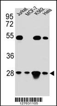
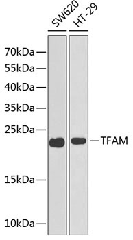
Western blot analysis in Hela, Jurkat, K562, MCF-7 cell line lysates (35 ug/lane).
Documents Download
Datasheet
Product Information
Request a Document
Protocol Information
WB
Western Blot (IB, immunoblot)
IHC-P
Immunohistochemistry Paraffin
FC
Flow Cytometry
IF
Immunofluorescence
TFAM Antibody (orb1270884)

- 0.0
Based on 0 reviews
Participating in our Biorbyt product reviews program enables you to support fellow scientists by sharing your firsthand experience with our products.
Login to Submit a ReviewAvailable Sizes
Select a size below
Free Secondary Antibody (20 ul)0/0
Please add an antibody product to your cart first.