You have no items in your shopping cart.
Description
Research Area
,Product Categories/Antibodies,Loading control
Images & Validation
−Item 1 of 3
| Tested Applications | IF, WB |
|---|---|
| Dilution Range | Western Blot 1:1000-1:3000 Immunofluorescence 1:200-1:500 |
| Reactivity | Human, Mouse, Rabbit |
Key Properties
−| Host | Rabbit |
|---|---|
| Clonality | Polyclonal |
| Isotype | Rabbit-IgG |
| Immunogen | Synthetic peptide / encompassing a sequence within the C-terminus region. |
| Target | Lamin B1 |
| Conjugation | Unconjugated |
Storage & Handling
−| Storage | Store at +4°C for short term storage. Long time storage is recommended at -20°C 100mM Tris Glycine, 1% BSA, 20% Glycerol (pH7). 0.025% ProClin 300 was added as a preservative |
|---|---|
| Expiration Date | 12 months from date of receipt. |
| Disclaimer | For research use only |
Similar Products
−- Item 1 of 11
Lamin B1/LMNB1 Antibody [orb312137]
FC, ICC, IF, IHC, IP, WB
Human, Mouse, Rat
Rabbit
Polyclonal
Unconjugated
100 μg - Item 1 of 9
- Item 1 of 9
Lamin B1 Antibody, KO Validated [orb1274472]
IF, IHC, WB
Human, Mouse, Rat
Polyclonal
Unconjugated
100 μl - Item 1 of 7
Lamin B1 Polyclonal Antibody [orb1413236]
IF, IHC-P, WB
Human, Mouse, Rat
Rabbit
Polyclonal
Unconjugated
100 μl - Item 1 of 7
Lamin B1 Recombinant Rabbit Monoclonal Antibody [orb704307]
ICC, IF, IHC-Fr, IHC-P, WB
Mouse, Rat
Human, Mouse, Rat
Rabbit
Recombinant
Unconjugated
100 μl, 50 μl


Quality Guarantee
Explore bioreagents carefree to elevate your research. All our products are rigorously tested for performance. If a product does not perform as described on its datasheet, our scientific support team will provide expert troubleshooting, a prompt replacement, or a refund. For full details, please see our Terms & Conditions and Buying Guide. Contact us at [email protected].

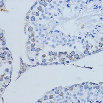
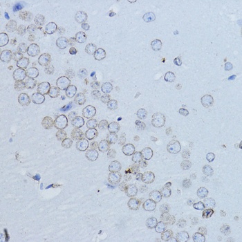
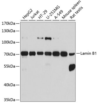
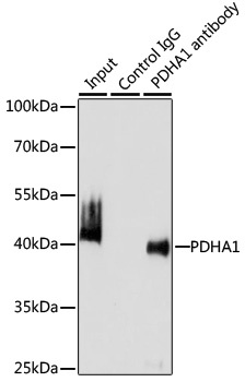
All lanes: Anti-Lamin B1 antibody - at 1/2000 dilution, Lysates/proteins at 50 μg per lane. This blot was produced using a 7.5% SDS-PAGE. The Nitrocellulose membrane was then blocked for an hour before being incubated with orb1294412 overnight at 4°C.

A549 cells'cytoplasmic and nuclear proteins extracted by 30-min. A. SDS-PAGE gel shows the profiles of extracted cytoplasmic proteins and nuclear proteins. B. The proteins from the gel were transferred to nitrocellulose membrane and probed with Lamin B1 antibody, Histone H3 antibody (nuclear marker) and GAPDH antibody (cytoplasmic marker). Nuclear Protein Extraction Kit.

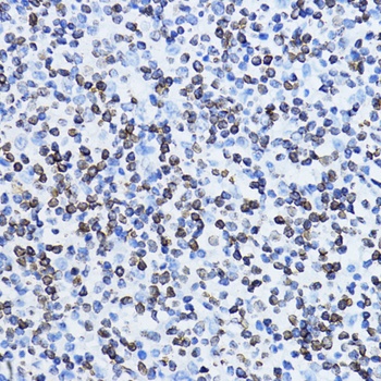
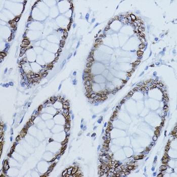
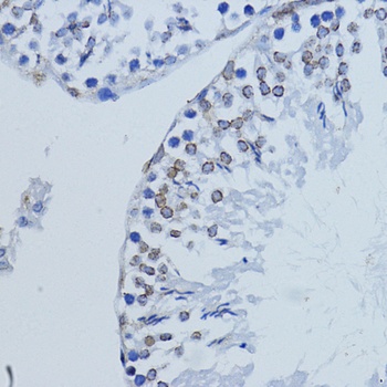
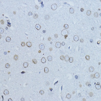
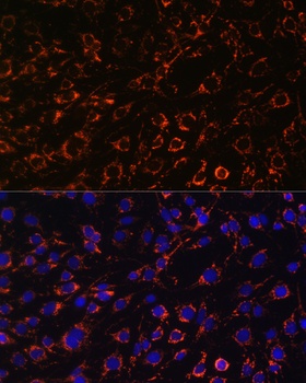
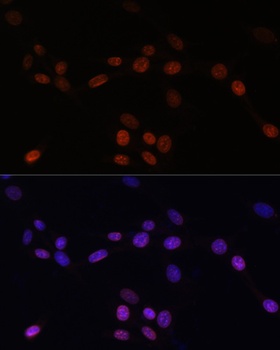
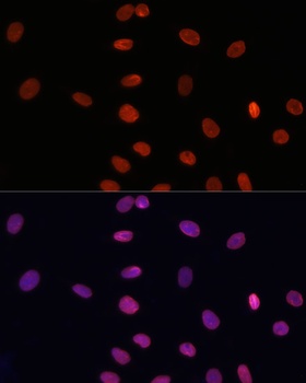
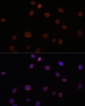
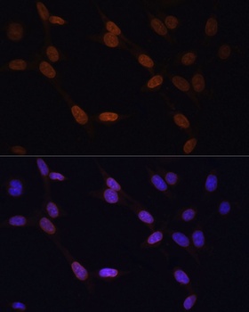
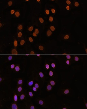
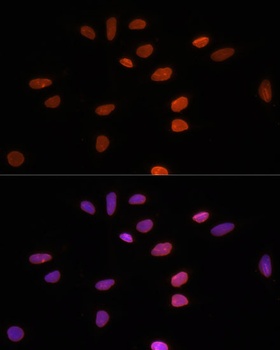
Immunofluorescence: cells were fixed with 4% paraformaldehyde for 10 min at RT, permeabilized with 0.1% NP-40 for 10 min at RT then blocked with 5% BSA for 30 min at room temperature. Cells were stained with orb1294412 anti-Lamin B1 antibody (red) at 1:200 and 4°C. DAPI (blue) was used as the nuclear counter stain.
Quick Database Links
Gene Symbol
Lamin B1
Documents Download
Datasheet
Product Information
Request a Document
Protocol Information
WB
Western Blot (IB, immunoblot)
IF
Immunofluorescence
Lamin B1 (orb1294412)

- 0.0
Based on 0 reviews
Participating in our Biorbyt product reviews program enables you to support fellow scientists by sharing your firsthand experience with our products.
Login to Submit a ReviewAvailable Sizes
Select a size below
Choose Conjugation or Carrier Free Version
Free Secondary Antibody (20 ul)0/0
Please add an antibody product to your cart first.