You have no items in your shopping cart.
Description
Research Area
Epigenetics, Infectious Diseases, Neuroscience
Images & Validation
−Item 1 of 5
| Tested Applications | FC, ICC, IHC-P, WB |
|---|---|
| Dilution Range | Immunocytochemistry: Staining technique: (a) fix cells for 10 min in methanol at -20°C and for 6 min in acetone at -20°C; (b) fix cells directly in methanol for 10 min at -20°C or in acetone for 10 min at -20°C. Positive control: 3T3 murine Swiss albino fibroblast cell line, RBL rat basophilic leukemia cell line. Flow cytometry: Recommended dilution: 1-5 µg/ml. Intracellular staining. Western blotting: Recommended dilution: 1 µg/ml. |
| Reactivity | Mammal |
| Application Notes |
Key Properties
−| Antibody Type | Primary Antibody |
|---|---|
| Clonality | Monoclonal |
| Isotype | Mouse IgM |
| Clone No. | VI-01 |
| Immunogen | Pellet of porcine brain cold stable proteins after depolymerization of microtubules. |
| Target | Vimentin |
| Purity | Purified by sequential steps of physicochemical fractionation (differential precipitation and solid-phase chromatography methods). |
| Purification | Purified by sequential steps of physicochemical fractionation (differential precipitation and solid-phase chromatography methods). |
| Conjugation | Unconjugated |
Storage & Handling
−| Storage | Maintain refrigerated at 2-8°C for up to 2 weeks. For long term storage store at -20°C in small aliquots to prevent freeze-thaw cycles. |
|---|---|
| Buffer/Preservatives | Tris buffered saline (TBS), pH 8.0, 15 mM sodium azide |
| Concentration | 1 mg/ml |
| Expiration Date | 12 months from date of receipt. |
| Disclaimer | For research use only |
Alternative Names
−anti CTRCT30 antibody, anti Epididymis luminal protein 113 antibody, anti HEL113 antibody, anti VIM antibody, anti Vimentin antibody
Similar Products
−- Item 1 of 5
Vimentin Antibody (Center) [orb34040]
FC, IHC-P, WB
Hamster, Mouse, Porcine, Rat
Human
Rabbit
Polyclonal
Unconjugated
80 μl - Item 1 of 4
Anti-Vimentin Purified [orb44572]
ICC, IHC-P, IP, WB
Gallus, Human, Mouse, Porcine, Rat
Monoclonal
Unconjugated
0.1 mg - Item 1 of 3
Anti-Vimentin Antibody [orb1821945]
ICC, WB
Gallus, Human, Mouse, Rat
Mouse
Monoclonal
Unconjugated
100 μlAnti-Vimentin Antibody [orb319708]
ICC, IHC, WB
Aves, Bovine, Canine, Drosophila, Equine, Feline, Fish, Gallus, Goat, Guinea pig, Hamster, Primate, Rabbit, Reptile, Rodent, Sheep, Xenopus, Zebrafish
Human, Mouse, Rat
Mouse
Monoclonal
Unconjugated
100 μl


Quality Guarantee
Explore bioreagents carefree to elevate your research. All our products are rigorously tested for performance. If a product does not perform as described on its datasheet, our scientific support team will provide expert troubleshooting, a prompt replacement, or a refund. For full details, please see our Terms & Conditions and Buying Guide. Contact us at [email protected].

Immunoprecipitation of vimentin from HeLa cell lysate by antibody VI-10 and its detection by antibody VI-01. IgM heavy chain (76-92 kDa) and IgM light chain (25-30 kDa) indicated. Mr of vimentin is 57 kDa. Lr = lysate (reducing conditions); Lnr = lysate (non-reducing conditions); IPr = immunoprecipitate (reducing conditions); IPnr = immunoprecipitate (non-reducing conditions).

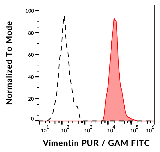
Separation of HeLa cells stained using anti-Vimentin (VI-01) purified antibody (concentration in sample 5 µg/ml, GAM APC, red-filled) from HeLa cells unstained by primary antibody (GAM APC, black-dashed) in flow cytometry analysis (intracellular staining of methanol permeabilisated cells).

Immunocytochemistry analysis of vimentin in methanol/acetone fixed murine 3T3 cells using mouse monoclonal antibody VI-01.

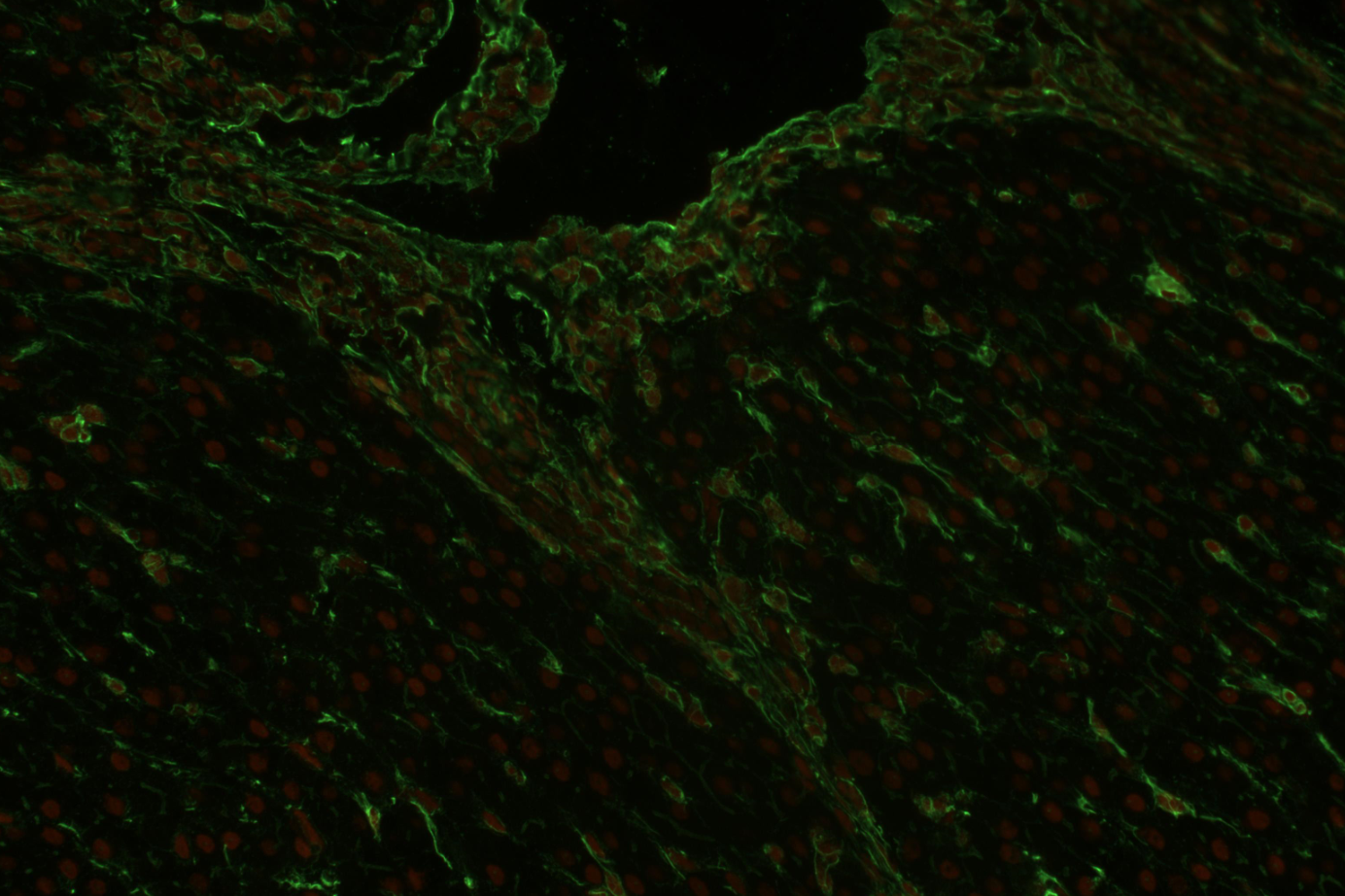
Immunohistochemistry staining (paraffin sections) of vimentin in human liver using mouse monoclonal antibody VI-01 (orb44570, diluted 1:400), detected with GAM IgM-Alexa Fluor®488 (diluted 1:200; green), cell nuclei stained with PI (1 µg/ml; orange).

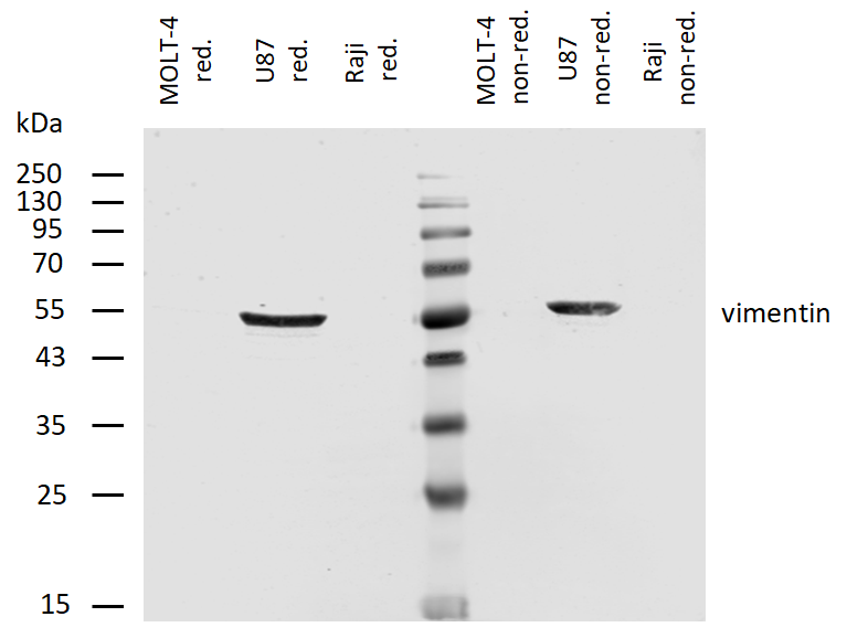
Anti-Vimentin Purified (VI-01) works in WB application under both reducing and non-reducing conditions. Western blotting analysis was performed on whole cell extracts (RIPA lysis buffer) of JAR, JEG3, HTr-8/SVneo, and HeLa cell lines mixed and heated (100°C, 5 min) with reducing (2-mercaptoethanol) or non-reducing SDS-loading buffer. Samples were resolved using 10% Tris-glycine SDS gel electrophoresis. Nitrocellulose membrane blot was probed simultaneously with mouse IgM monoclonal antibody VI-01 (1 µg/ml), and mouse IgG1 anti-tubulin monoclonal antibody TU-01 (1 µg/ml) used as the loading control. Subclass-specific secondary antibodies IRDye 800CW Goat-anti-Mouse IgG (green) and IRDye 680RD Goat-anti-Mouse IgM (red) were used for multiplex fluorescent Western blot detection. Vimentin was detected at ~55 kDa.
Documents Download
Datasheet
Product Information
Request a Document
Protocol Information
WB
Western Blot (IB, immunoblot)
IHC-P
Immunohistochemistry Paraffin
FC
Flow Cytometry
ICC
Immunocytochemistry
Anti-Vimentin Purified (orb44570)

- 0.0
Based on 0 reviews
Participating in our Biorbyt product reviews program enables you to support fellow scientists by sharing your firsthand experience with our products.
Login to Submit a ReviewAvailable Sizes
Select a size below
Free Secondary Antibody (20 ul)0/0
Please add an antibody product to your cart first.